2.8 The Nucleus and DNA Replication
Learning Objectives
By the end of this section, you will be able to:
- Describe the structure and features of the nuclear membrane
- List the contents of the nucleus
- Explain the organisation of the DNA molecule within the nucleus
- Describe the process of DNA replication
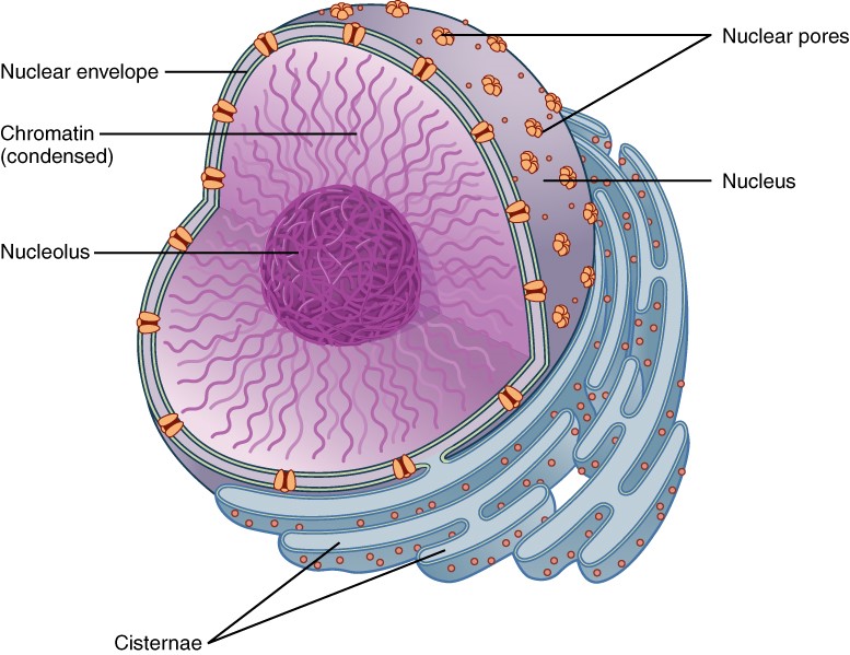
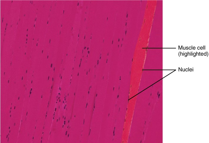
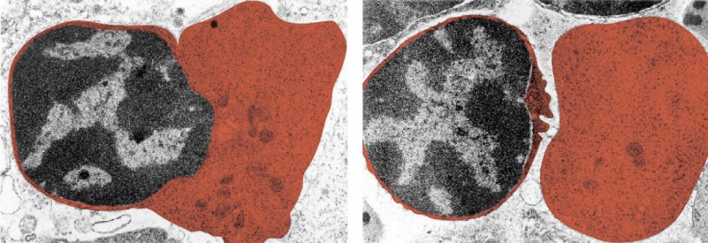
The nucleus is the largest and most prominent of a cell’s organelles (Figure 2.8.1). The nucleus is generally considered the control centre of the cell because it stores all of the genetic instructions for manufacturing proteins. Interestingly, some cells in the body, such as muscle cells, contain more than one nucleus (Figure 2.8.2), which is known as multinucleated. Other cells, such as mammalian red blood cells (RBCs), do not contain nuclei at all. RBCs eject their nuclei as they mature, making space for the large numbers of haemoglobin molecules that carry oxygen throughout the body (Figure 2.8.3). Without nuclei, the life span of RBCs is short, and so the body must produce new ones constantly.
Inside the nucleus lies the blueprint that dictates everything a cell will do and all the products it will make. This information is stored within DNA. The nucleus sends “commands” to the cell via molecular messengers that translate the information from DNA. Each cell in your body (with the exception of germ cells) contains the complete set of your DNA. When a cell divides, the DNA must be duplicated so that each new cell receives a full complement of DNA. The following section will explore the structure of the nucleus and its contents, as well as the process of DNA replication.
Organisation of the Nucleus and its DNA
Like most other cellular organelles, the nucleus is surrounded by a membrane called the nuclear envelope. This membranous covering consists of two adjacent lipid bilayers with a thin fluid space in between them. Spanning these two bilayers are nuclear pores. A nuclear pore is a tiny passageway for the passage of proteins, RNA, and solutes between the nucleus and the cytoplasm. Proteins called pore complexes lining the nuclear pores regulate the passage of materials into and out of the nucleus.
Inside the nuclear envelope is a gel-like nucleoplasm with solutes that include the building blocks of nucleic acids. There also can be a dark-staining mass often visible under a simple light microscope, called a nucleolus (plural = nucleoli). The nucleolus is a region of the nucleus that is responsible for manufacturing the RNA necessary for construction of ribosomes. Once synthesised, newly made ribosomal subunits exit the cell’s nucleus through the nuclear pores.
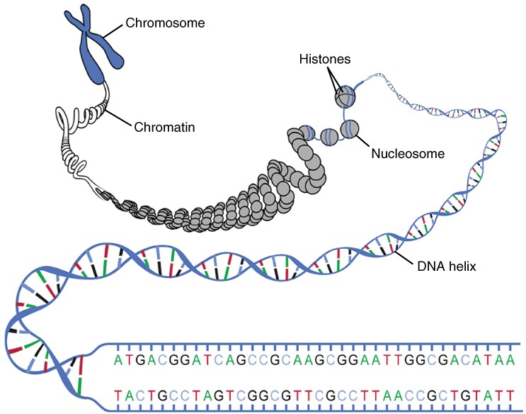
The genetic instructions that are used to build and maintain an organism are arranged in an orderly manner in strands of DNA. Within the nucleus are threads of chromatin composed of DNA and associated proteins (Figure 2.8.4). Along the chromatin threads, the DNA is wrapped around a set of histone proteins. A nucleosome is a single, wrapped DNA-histone complex. Multiple nucleosomes along the entire molecule of DNA appear like a beaded necklace, in which the string is the DNA and the beads are the associated histones. When a cell is in the process of division, the chromatin condenses into chromosomes, so that the DNA can be safely transported to the “daughter cells.” The chromosome is composed of DNA and proteins; it is the condensed form of chromatin. It is estimated that humans have almost 22,000 genes distributed on 46 chromosomes.
DNA Replication
In order for an organism to grow, develop, and maintain its health, cells must reproduce themselves by dividing to produce two new daughter cells, each with the full complement of DNA as found in the original cell. Billions of new cells are produced in an adult human every day. Only very few cell types in the body do not divide, including nerve cells, skeletal muscle fibres and cardiac muscle cells. The division time of different cell types varies. Epithelial cells of the skin and gastrointestinal lining, for instance, divide very frequently to replace those that are constantly being rubbed off the surface by friction.
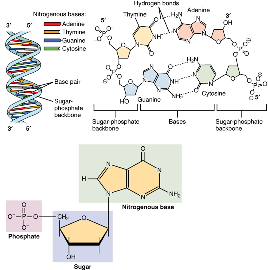
A DNA molecule is made of two strands that “complement” each other in the sense that the molecules that compose the strands fit together and bind to each other, creating a double-stranded molecule that looks much like a long, twisted ladder. Each side rail of the DNA ladder is composed of alternating sugar and phosphate groups (Figure 2.8.5). The two sides of the ladder are not identical, but are complementary. These two backbones are bonded to each other across pairs of protruding bases, each bonded pair forming one “rung,” or cross member. The four DNA bases are adenine (A), thymine (T), cytosine (C), and guanine (G). Because of their shape and charge, the two bases that compose a pair always bond together. Adenine always binds with thymine, and cytosine always binds with guanine. The particular sequence of bases along the DNA molecule determines the genetic code. Therefore, if the two complementary strands of DNA were pulled apart, you could infer the order of the bases in one strand from the bases in the other, complementary strand. For example, if one strand has a region with the sequence AGTGCCT, then the sequence of the complementary strand would be TCACGGA.
DNA replication is the copying of DNA that occurs before cell division can take place. After a great deal of debate and experimentation, the general method of DNA replication was deduced in 1958 by two scientists in California, Matthew Meselson and Franklin Stahl. This method is illustrated in Figure 2.8.6 and described below.
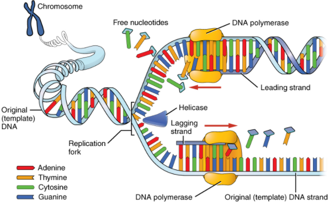
Stage 1: Initiation. The two complementary strands are separated, much like unzipping a zipper. Special enzymes, including helicase, untwist and separate the two strands of DNA.
Stage 2: Elongation. Each strand becomes a template along which a new complementary strand is built. DNA polymerase brings in the correct bases to complement the template strand, synthesising a new strand base by base. A DNA polymerase is an enzyme that adds free nucleotides to the end of a chain of DNA, making a new double strand. This growing strand continues to be built until it has fully complemented the template strand.
Stage 3: Termination. Once the two original strands are bound to their own, finished, complementary strands, DNA replication is stopped and the two new identical DNA molecules are complete.
Each new DNA molecule contains one strand from the original molecule and one newly synthesised strand. The term for this mode of replication is “semiconservative,” because half of the original DNA molecule is conserved in each new DNA molecule. This process continues until the cell’s entire genome, the entire complement of an organism’s DNA, is replicated. As you might imagine, it is very important that DNA replication take place precisely so that new cells in the body contain the exact same genetic material as their parent cells. Mistakes made during DNA replication, such as the accidental addition of an inappropriate nucleotide, have the potential to render a gene dysfunctional or useless. Fortunately, there are mechanisms in place to minimise such mistakes. A DNA proofreading process enlists the help of special enzymes that scan the newly synthesised molecule for mistakes and corrects them. Once the process of DNA replication is complete, the cell is ready to divide. You will explore the process of cell division later in the chapter.
Section Review
The nucleus is the command centre of the cell, containing the genetic instructions for all the materials a cell will make (and thus all of its functions it can perform). The nucleus is encased within a membrane of two interconnected lipid bilayers, side-by-side. This nuclear envelope is studded with protein-lined pores that allow materials to be trafficked into and out of the nucleus. The nucleus contains one or more nucleoli, which serve as sites for ribosome synthesis. The nucleus houses the genetic material of the cell: DNA. DNA is normally found as a loosely contained structure called chromatin within the nucleus, where it is wound up and associated with a variety of histone proteins. When a cell is about to divide, the chromatin coils tightly and condenses to form chromosomes.
There is a pool of cells constantly dividing within your body. The result is billions of new cells being created each day. Before any cell is ready to divide, it must replicate its DNA so that each new daughter cell will receive an exact copy of the organism’s genome. A variety of enzymes are enlisted during DNA replication. These enzymes unwind the DNA molecule, separate the two strands, and assist with the building of complementary strands along each parent strand. The original DNA strands serve as templates from which the nucleotide sequence of the new strands are determined and synthesised. When replication is completed, two identical DNA molecules exist. Each one contains one original strand and one newly synthesised complementary strand.
Review Questions
Critical Thinking Questions
Click the drop down below to review the terms learned from this chapter.
