16.3 Foetal Development
Learning Objectives
By the end of this section, you will be able to:
- Differentiate between the embryonic period and the foetal period
- Briefly describe the process of sexual differentiation
- Describe the foetal circulatory system and explain the role of the shunts
- Trace the development of a foetus from the end of the embryonic period to birth
As you will recall, a developing human is called a foetus from the ninth week of gestation until birth. This 30-week period of development is marked by continued cell growth and differentiation, which fully develop the structures and functions of the immature organ systems formed during the embryonic period. The completion of foetal development results in a newborn who, although still immature in many ways, is capable of survival outside the womb.
Sexual Differentiation
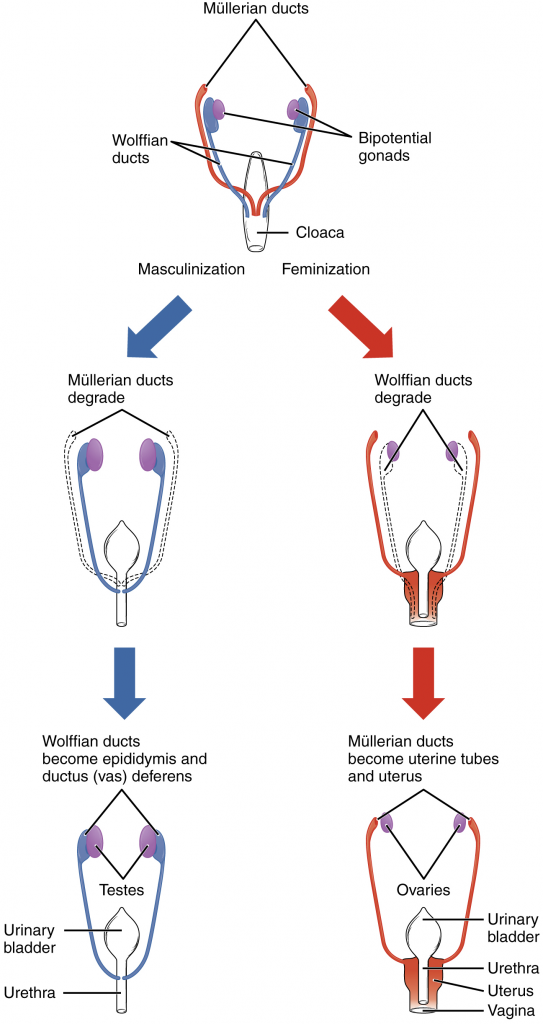
Sexual differentiation does not begin until the foetal period, during weeks 9–12. Embryonic males (with SRY gene) and females, though genetically distinguishable, are morphologically identical (Figure 16.3.1). Bipotential gonads, or gonads that can develop into male or female sexual organs, are connected to a central cavity called the cloaca via Müllerian ducts and Wolffian ducts. (The cloaca is an extension of the primitive gut.) Several events lead to sexual differentiation during this period.
During male foetal development, the bipotential gonads become the testes and associated epididymis. The Müllerian ducts degenerate. The Wolffian ducts become the vas deferens and the cloaca becomes the urethra and rectum.
During female foetal development, the bipotential gonads develop into ovaries. The Wolffian ducts degenerate. The Müllerian ducts become the uterine tubes and uterus and the cloaca divides and develops into a vagina, a urethra and a rectum.
The Foetal Circulatory System
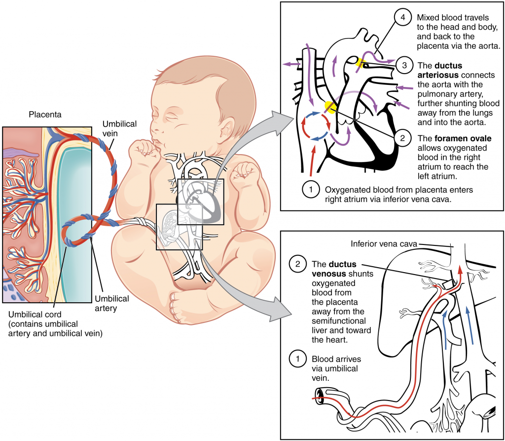
During prenatal development, the foetal circulatory system is integrated with the placenta via the umbilical cord so that the foetus receives both oxygen and nutrients from the placenta. However, after childbirth, the umbilical cord is severed, and the newborn’s circulatory system must be reconfigured. When the heart first forms in the embryo, it exists as two parallel tubes derived from mesoderm and lined with endothelium, which then fuse together. As the embryo develops into a foetus, the tube-shaped heart folds and further differentiates into the four chambers present in a mature heart. Unlike a mature cardiovascular system, however, the foetal cardiovascular system also includes circulatory shortcuts, or shunts. A shunt is an anatomical (or sometimes surgical) diversion that allows blood flow to bypass immature organs such as the lungs and liver until childbirth.
The placenta provides the foetus with necessary oxygen and nutrients via the umbilical vein. (Remember that veins carry blood toward the heart. In this case however, the blood flowing to the foetal heart is oxygenated because it comes from the placenta. The respiratory system is immature and cannot yet oxygenate blood on its own.) From the umbilical vein, the oxygenated blood flows toward the inferior vena cava, all but bypassing the immature liver, via the ductus venosus shunt (Figure 16.3.2). The liver receives just a trickle of blood, which is all that it needs in its immature, semifunctional state. Blood flows from the inferior vena cava to the right atrium, mixing with foetal venous blood along the way.
Although the foetal liver is semifunctional, the foetal lungs are non-functional. The foetal circulation therefore bypasses the lungs by shifting some of the blood through the foramen ovale, a shunt that directly connects the right and left atria and avoids the pulmonary trunk altogether. Most of the rest of the blood is pumped to the right ventricle, and from there, into the pulmonary trunk, which splits into pulmonary arteries. However, a shunt within the pulmonary artery, the ductus arteriosus, diverts a portion of this blood into the aorta. This ensures that only a small volume of oxygenated blood passes through the immature pulmonary circuit, which has only minor metabolic requirements. Blood vessels of uninflated lungs have high resistance to flow, a condition that encourages blood to flow to the aorta, which presents much lower resistance. The oxygenated blood moves through the foramen ovale into the left atrium, where it mixes with the now deoxygenated blood returning from the pulmonary circuit. This blood then moves into the left ventricle, where it is pumped into the aorta. Some of this blood moves through the coronary arteries into the myocardium and some moves through the carotid arteries to the brain.
The descending aorta carries partially oxygenated and partially deoxygenated blood into the lower regions of the body. It eventually passes into the umbilical arteries through branches of the internal iliac arteries. The deoxygenated blood collects waste as it circulates through the foetal body and returns to the umbilical cord. Thus, the two umbilical arteries carry blood low in oxygen and high in carbon dioxide and foetal wastes. This blood is filtered through the placenta, where wastes diffuse into the maternal circulation. Oxygen and nutrients from the mother diffuse into the placenta and from there into the foetal blood, and the process repeats.
Other Organ Systems
During weeks 9–12 of foetal development, the brain continues to expand, the body elongates, and ossification continues. Foetal movements are frequent during this period but are jerky and not well-controlled. The bone marrow begins to take over the process of erythrocyte production—a task that the liver performed during the embryonic period. The liver now secretes bile. The foetus circulates amniotic fluid by swallowing it and producing urine. The eyes are well-developed by this stage, but the eyelids are fused shut. The fingers and toes begin to develop nails. By the end of week 12, the foetus measures approximately 9 cm from crown to rump.
Weeks 13–16 are marked by sensory organ development. The eyes move closer together; blinking motions begin, although the eyes remain sealed shut. The lips exhibit sucking motions. The ears move upward and lie flatter against the head. The scalp begins to grow hair. The excretory system is also developing: the kidneys are well-formed, and meconium, or foetal faeces, begins to accumulate in the intestines. Meconium consists of ingested amniotic fluid, cellular debris, mucus and bile.
During approximately weeks 16–20, as the foetus grows and limb movements become more powerful, the mother may begin to feel quickening, or foetal movements. However, space restrictions limit these movements and typically force the growing foetus into the “foetal position,” with the arms crossed and the legs bent at the knees. Sebaceous glands coat the skin with a waxy, protective substance called vernix caseosa that protects and moisturises the skin and may provide lubrication during childbirth. A silky hair called lanugo also covers the skin during weeks 17–20, but it is shed as the foetus continues to grow. Extremely premature infants sometimes exhibit residual lanugo.
Developmental weeks 21–30 are characterised by rapid weight gain, which is important for maintaining a stable body temperature after birth. The bone marrow completely takes over erythrocyte synthesis, and the axons of the spinal cord begin to be myelinated or coated in the electrically insulating glial cell sheaths that are necessary for efficient nervous system functioning. (The process of myelination is not completed until adolescence.) During this period, the foetus grows eyelashes. The eyelids are no longer fused and can be opened and closed. The lungs begin producing surfactant, a substance that reduces surface tension in the lungs and assists proper lung expansion after birth. Inadequate surfactant production in premature newborns may result in respiratory distress syndrome, and as a result, the newborn may require surfactant replacement therapy, supplemental oxygen, or maintenance in a continuous positive airway pressure (CPAP) chamber during their first days or weeks of life. In male foetuses, the testes descend into the scrotum near the end of this period. The foetus at 30 weeks measures 28 cm from crown to rump and exhibits the approximate body proportions of a full-term newborn, but still is much leaner.
The foetus continues to lay down subcutaneous fat from week 31 until birth. The added fat fills out the hypodermis and the skin transitions from red and wrinkled to soft and pink. Lanugo is shed, and the nails grow to the tips of the fingers and toes. Immediately before birth, the average crown-to-rump length is 35.5–40.5 cm, and the foetus weighs approximately 2.5–4 kg. Once born, the newborn is no longer confined to the foetal position, so subsequent measurements are made from head-to-toe instead of from crown-to-rump. At birth, the average length is approximately 51 cm.
Disorders of the Developing Foetus
Throughout the second half of gestation, the foetal intestines accumulate a tarry, greenish black meconium. The newborn’s first stools consist entirely of meconium; they later transition to seedy yellow stools or slightly formed tan stools as meconium is cleared and replaced with digested breast milk or formula, respectively. Unlike these later stools, meconium is sterile; it is devoid of bacteria because the foetus is in a sterile environment and has not consumed any breast milk or formula. Typically, an infant does not pass meconium until after birth. However, in 5–20 percent of births, the foetus has a bowel movement in utero, which can cause major complications in the newborn.
The passage of meconium in the uterus signals foetal distress, particularly foetal hypoxia (i.e., oxygen deprivation). This may be caused by maternal drug abuse (especially tobacco or cocaine), maternal hypertension, depletion of amniotic fluid, long labour or difficult birth, or a defect in the placenta that prevents it from delivering adequate oxygen to the foetus. Meconium passage is typically a complication of full-term or post-term newborns because it is rarely passed before 34 weeks of gestation, when the gastrointestinal system has matured and is appropriately controlled by nervous system stimuli. Foetal distress can stimulate the vagus nerve to trigger gastrointestinal peristalsis and relaxation of the anal sphincter. Notably, foetal hypoxic stress also induces a gasping reflex, increasing the likelihood that meconium will be inhaled into the foetal lungs.
Although meconium may be considered a sterile substance, it interferes with the antibiotic properties of the amniotic fluid and makes the newborn and mother more vulnerable to bacterial infections at birth and during the perinatal period. Specifically, inflammation of the foetal membranes, inflammation of the uterine lining, or neonatal sepsis (infection in the newborn) may occur. Meconium also irritates delicate foetal skin and can cause a rash.
The first sign that a foetus has passed meconium usually does not come until childbirth, when the amniotic sac ruptures. Normal amniotic fluid is clear and watery, but amniotic fluid in which meconium has been passed is stained greenish or yellowish. Antibiotics given to the mother may reduce the incidence of maternal bacterial infections, but it is critical that meconium is aspirated from the newborn before the first breath. Under these conditions, an obstetrician will extensively aspirate the infant’s airways as soon as the head is delivered, while the rest of the infant’s body is still inside the birth canal.
Aspiration of meconium with the first breath can result in laboured breathing, a barrel-shaped chest, or a low Apgar score. An obstetrician can identify meconium aspiration by listening to the lungs with a stethoscope for a coarse rattling sound. Blood gas tests and chest X-rays of the infant can confirm meconium aspiration. Inhaled meconium after birth could obstruct a newborn’s airways leading to alveolar collapse, interfere with surfactant function by stripping it from the lungs or cause pulmonary inflammation or hypertension. Any of these complications will make the newborn much more vulnerable to pulmonary infection, including pneumonia.
Section Review
The foetal period lasts from the ninth week of development until birth. During this period, male and female gonads differentiate. The foetal circulatory system becomes much more specialised and efficient than its embryonic counterpart. It includes three shunts—the ductus venosus, the foramen ovale and the ductus arteriosus—that enable it to bypass the semifunctional liver and pulmonary circuit until after childbirth. The brain continues to grow and its structures differentiate. Facial features develop, the body elongates and the skeleton ossifies. In the womb, the developing foetus moves, blinks, practices sucking and circulates amniotic fluid. The foetus grows from an embryo measuring approximately 3.3 cm and weighing 7 g to an infant measuring approximately 51 cm and weighing an average of approximately 3.4 kg. Embryonic organ structures that were primitive and non-functional develop to the point that the newborn can survive in the outside world.
Review Questions
Critical Thinking Questions
Click the drop down below to review the terms learned from this chapter.
