13 Eukaryote Cell Cycle
In this section
Content in this section includes:
Within the field of developmental biology in humans, one goal is to understand how a particular cell develops into a final cell type; this is referred to as fate determination. For a cell to arrive at its final cell type many processes are undertaken including cell proliferation (growth and division), differentiation, cellular movement and programmed cell death or apoptosis. There are many processes, checks and balances to ensure the cell cycles proceed as precisely and smoothly are possible and for apoptosis to occur when needed and appropriate.
The cell cycle is a highly ordered series of events involving cell growth and cell division that produces two new daughter cells. This mitotic replication happens to almost every cell in the body, except for sex cells or gametes. Gametes replicate through a slightly different process called meiosis. Mitotic cell division involves a series of precisely timed and carefully regulated stages of growth, DNA replication and division that produce two genetically identical cells. The cell cycle has two major phases: interphase and the mitotic phase (Figure 4.4 and Figure 4.5). During interphase, the cell grows and the DNA is replicated. During the mitotic phase, the replicated DNA and cytoplasmic contents are separated and the cell divides.
The cell cycle is completed in about 24 hours in most human cells however some cell types have slower or faster cell cycle times and there are a number of controls and checkpoints to ensure the cell cycle is completed correctly.
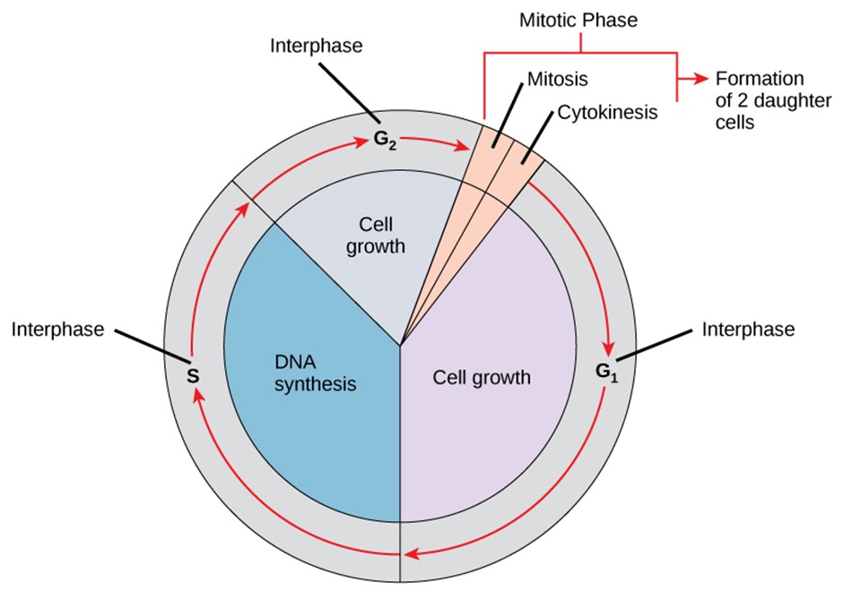
Interphase
During interphase, the cell undergoes normal processes while also preparing for cell division. For a cell to move from interphase to the mitotic phase, many internal and external conditions must be met. The three stages of interphase are called the first gap (G1), phase of DNA synthesis (S phase), and the second gap (G2). During interphase, there are no visible changes to the cell’s morphology.
Interphase: G1 phase
The first stage of interphase is called the G1 phase, or first gap, because little change is visible. However, during the G1 stage, the cell is quite active at the biochemical level. The cell is accumulating the building blocks of chromosomal DNA and the associated proteins, as well as accumulating enough energy reserves to complete the task of replicating each chromosome in the nucleus. This is the longer of the gap phases, immediately following cell division and is directly responsible for cell fate determination.
Interphase: S phase
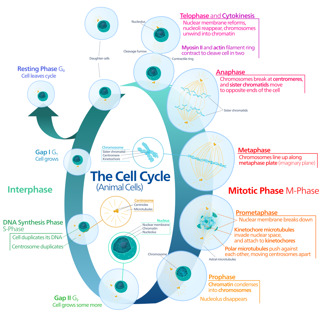
Throughout interphase, nuclear DNA remains in a semi-condensed chromatin configuration. In the S phase (synthesis phase), DNA replication results in the formation of two identical copies of each chromosome—sister chromatids—that are firmly attached at the centromere region. At this stage, each chromosome is made of two sister chromatids and is a duplicated chromosome. The centrosome is duplicated during the S phase. The two centrosomes will give rise to the mitotic spindle, the apparatus that orchestrates the movement of chromosomes during mitosis. The centrosome consists of a pair of rod-like centrioles at right angles to each other. Centrioles help organise cell division. Centrioles are not present in the centrosomes of many eukaryotic species, such as plants and most fungi.
Interphase: G2 phase
In the G2 phase, or second gap, the cell replenishes its energy stores and synthesises the proteins necessary for chromosome manipulation. Some cell organelles are duplicated, and the cytoskeleton is dismantled to provide resources for the mitotic spindle. There may be additional cell growth during G2. The final preparations for the mitotic phase must be completed before the cell is able to enter the first stage of mitosis.
Mitotic Phase
To make two daughter cells, the contents of the nucleus and the cytoplasm must be divided. The mitotic phase is a multistep process during which the duplicated chromosomes are aligned, separated, and moved to opposite poles of the cell, and then the cell is divided into two new identical daughter cells. The first portion of the mitotic phase, mitosis (or karyokinesis), is composed of five stages, which accomplish nuclear division. The second portion of the mitotic phase, called cytokinesis, is the physical separation of the cytoplasmic components into two daughter cells.
Mitotic Phase: Mitosis
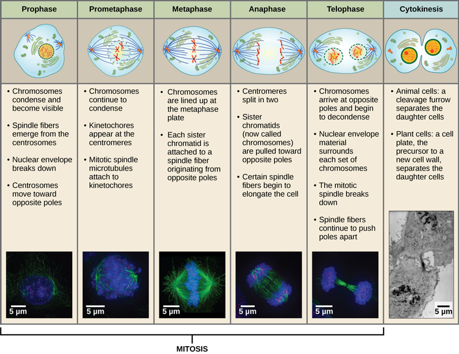
Mitosis is divided into a series of phases—prophase, prometaphase, metaphase, anaphase and telophase—that result in the division of the cell nucleus (Figure 4.6).
Prophase
Prophase (the “first phase”): the nuclear envelope starts to dissociate into small vesicles, and the membranous organelles (such as the Golgi complex [Golgi apparatus] and the endoplasmic reticulum), fragment and disperse toward the periphery of the cell. The nucleolus disappears (disperses) as well, and the centrosomes begin to move to opposite poles of the cell. Microtubules that will form the mitotic spindle extend between the centrosomes, pushing them farther apart as the microtubule fibres lengthen. The sister chromatids begin to coil more tightly with the aid of condensin proteins and now become visible under a light microscope.
Prometaphase
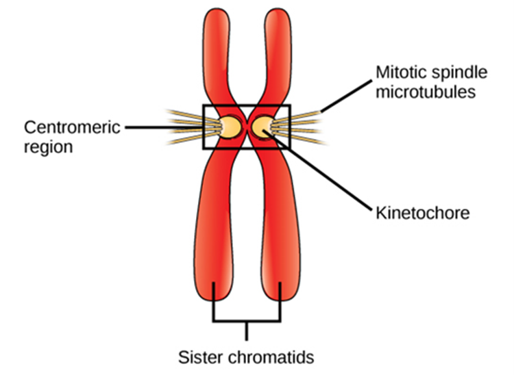
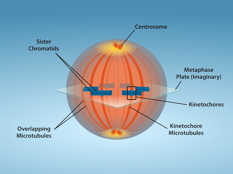
Prometaphase (the “first change phase”): Many processes that began in prophase continue to advance. The remnants of the nuclear envelope fragment further, and the mitotic spindle continues to develop as more microtubules assemble and stretch across the length of the former nuclear area. Chromosomes become even more condensed and discrete. Each sister chromatid develops a protein structure called a kinetochore in its centromeric region (Figure 4.7). The proteins of the kinetochore attract and bind to the mitotic spindle microtubules. As the spindle microtubules extend from the centrosomes, some of these microtubules come into contact with and firmly bind to the kinetochores. Once a mitotic fibre attaches to a chromosome, the chromosome will be oriented until the kinetochores of sister chromatids face the opposite poles. Eventually, all the sister chromatids will be attached via their kinetochores to microtubules from opposing poles. Spindle microtubules that do not engage the chromosomes are called polar microtubules. These microtubules overlap each other midway between the two poles and contribute to cell elongation. Astral microtubules are located near the poles, aid in spindle orientation, and are required for the regulation of mitosis.
Metaphase
Metaphase (the “change phase”): All the chromosomes are aligned in a plane called the metaphase plate, or the equatorial plane, roughly midway between the two poles of the cell. The sister chromatids are still tightly attached to each other by special cohesin proteins (Figure 4.8). At this time, the chromosomes are maximally condensed and this is the stage that cells are used in karyotyping.
Anaphase
Anaphase (“upward phase”): The cohesin proteins degrade and the sister chromatids separate at the centromere. Each chromatid, now called a single chromosome, is pulled rapidly toward the centrosome to which its microtubule is attached. The cell becomes visibly elongated (oval shaped) as the polar microtubules slide against each other at the metaphase plate where they overlap.
Telophase
During telophase (the “distance phase”): all of the events that set up the duplicated chromosomes for mitosis during the first three phases are reversed. The chromosomes reach the opposite poles and begin to decondense (unravel), relaxing once again into a stretched-out chromatin configuration. The mitotic spindles are depolymerised into tubulin monomers that will be used to assemble cytoskeletal components for each daughter cell. Nuclear envelopes form around the chromosomes, and nucleosomes appear within the nuclear area.
Mitotic phase: Cytokinesis
Cytokinesis is the second part of the mitotic phase during which cell division is completed by the physical separation of the cytoplasmic components into two daughter cells. Although the stages of mitosis are similar for most eukaryotes, the process of cytokinesis is quite different for eukaryotes that have cell walls, such as plant cells.
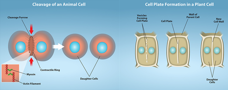
In cells such as animal cells that lack cell walls, cytokinesis begins following the onset of anaphase. A contractile ring composed of actin filaments forms just inside the plasma membrane at the former metaphase plate. The actin filaments pull the equator of the cell inward, forming a fissure. This fissure, or “crack,” is called the cleavage furrow. The furrow deepens as the actin ring contracts, and eventually the membrane and cell are cleaved in two (Figure 4.9).
In plant cells, a cleavage furrow is not possible because of the rigid cell walls surrounding the plasma membrane. A new cell wall must form between the daughter cells. Therefore, during interphase, the Golgi apparatus accumulates enzymes, structural proteins and glucose molecules prior to breaking up into vesicles and dispersing throughout the dividing cell. During telophase, these Golgi vesicles move on microtubules to collect at the metaphase plate. There, the vesicles fuse from the centre toward the cell walls; this structure is called a cell plate. As more vesicles fuse, the cell plate enlarges until it merges with the cell wall at the periphery of the cell. Enzymes use the glucose that has accumulated between the membrane layers to build a new cell wall of cellulose. The Golgi membranes become the plasma membrane on either side of the new cell wall (Figure 4.9).
Meiosis (for Sexual Reproduction)
Sexual reproduction requires fertilisation, a union of two cells from two individual organisms. If those two cells each contain one set of chromosomes, then the resulting cell contains two sets of chromosomes. The number of sets of chromosomes in a cell is called its ploidy level. Haploid cells contain one set of chromosomes. Cells containing two sets of chromosomes are called diploid. If the reproductive cycle is to continue, the diploid cell must somehow reduce its number of chromosome sets before fertilisation can occur again or there will be a continual doubling in the number of chromosome sets in every generation. So, in addition to fertilisation, sexual reproduction includes a nuclear division, known as meiosis, that reduces the number of chromosome sets.
The nuclear division that forms haploid cells, which is called meiosis, is related to mitosis. In mitosis, both the parent and the daughter nuclei contain the same number of chromosome sets—diploid for most plants and animals. Meiosis employs many of the same mechanisms as mitosis. However, the starting nucleus is always diploid and the nuclei that result at the end of a meiotic cell division are haploid. To achieve the reduction in chromosome number, meiosis consists of one round of chromosome duplication and two rounds of nuclear division. Because the events that occur during each of the division stages are analogous to the events of mitosis, the same stage names are assigned. However, because there are two rounds of division, the stages are designated with a “I” or “II.” Thus, meiosis I is the first round of meiotic division and consists of prophase I, prometaphase I, and so on. Meiosis I reduces the number of chromosome sets from two to one. The genetic information is also mixed during this division to create unique recombinant chromosomes. Meiosis II, in which the second round of meiotic division takes place in a way that is like mitosis, includes prophase II, prometaphase II, and so on.
Interphase
Meiosis is preceded by an interphase consisting of the G1, S, and G2 phases, which are nearly identical to the phases preceding mitosis. The G1 phase is the first phase of interphase and is focused on cell growth. In the S phase, the DNA of the chromosomes is replicated. Finally, in the G2 phase, the cell undergoes the final preparations for meiosis.
During DNA duplication of the S phase, each chromosome becomes composed of two identical copies (called sister chromatids) that are held together at the centromere until they are pulled apart during meiosis II. In an animal cell, the centrosomes that organise the microtubules of the meiotic spindle also replicate. This prepares the cell for the first meiotic phase.
Meiosis I
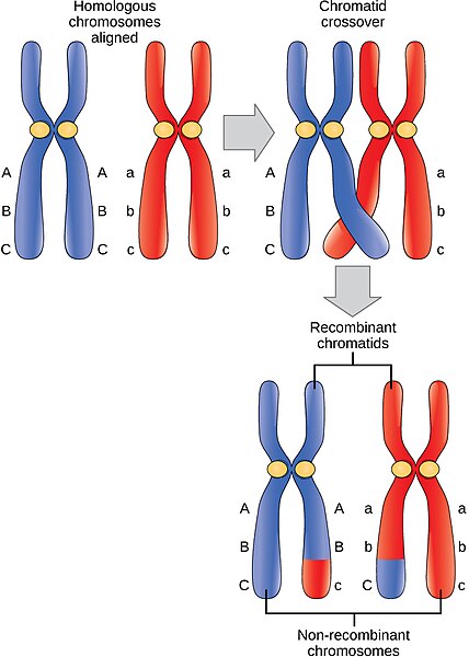
Early in prophase I, the chromosomes can be clearly seen microscopically. As the nuclear envelope begins to break down, the proteins associated with homologous chromosomes bring the pair close to each other. The tight pairing of the homologous chromosomes is called synapsis. In synapsis, the genes on the chromatids of the homologous chromosomes are precisely aligned with each other. An exchange of chromosome segments between non-sister homologous chromatids occurs and is called crossing over. This process is revealed visually after the exchange as chiasmata (singular = chiasma) (Figure 4.10).
As prophase I progresses, the close association between homologous chromosomes begins to break down, and the chromosomes continue to condense, although the homologous chromosomes remain attached to each other at chiasmata. The number of chiasmata varies with the species and the length of the chromosome. At the end of prophase I, the pairs are held together only at chiasmata (Figure 4.10) and are called tetrads because the four sister chromatids of each pair of homologous chromosomes are now visible.
The crossover events are the first source of genetic variation produced by meiosis. A single crossover event between homologous non-sister chromatids leads to a reciprocal exchange of equivalent DNA between a maternal chromosome and a paternal chromosome. Now, when that sister chromatid is moved into a gamete, it will carry some DNA from one parent of the individual and some DNA from the other parent. The recombinant sister chromatid has a combination of maternal and paternal genes that did not exist before the crossover.
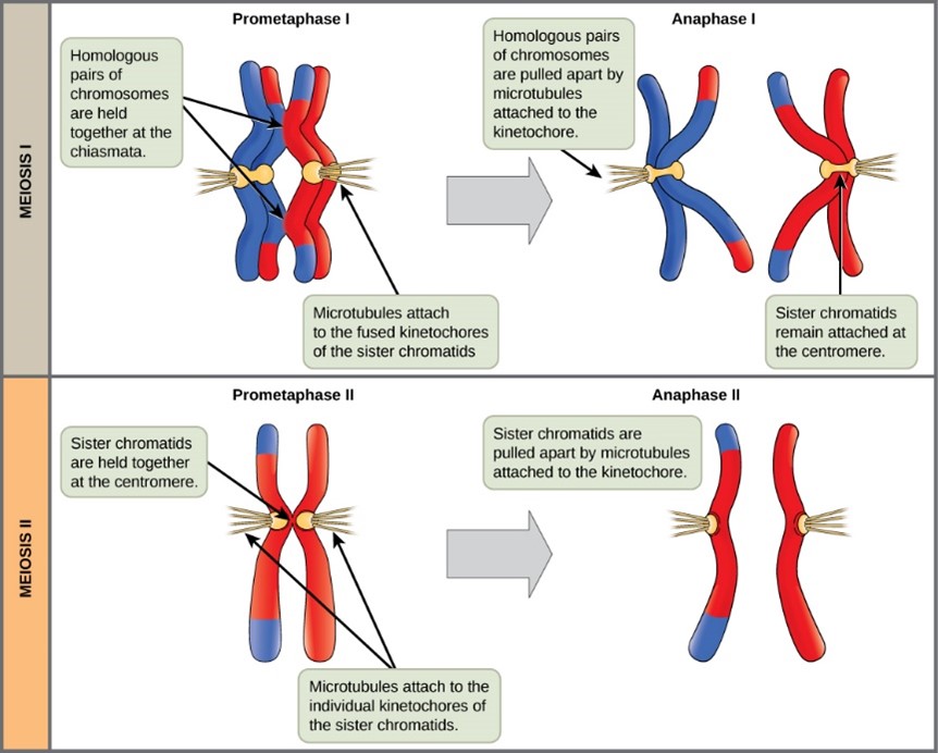
The key event in prometaphase I is the attachment of the spindle fibre microtubules to the kinetochore proteins at the centromeres. The microtubules assembled from centrosomes at opposite poles of the cell grow toward the middle of the cell. At the end of prometaphase I, each tetrad is attached to microtubules from both poles, with one homologous chromosome attached at one pole and the other homologous chromosome attached to the other pole. The homologous chromosomes are still held together at chiasmata. In addition, the nuclear membrane has broken down entirely.
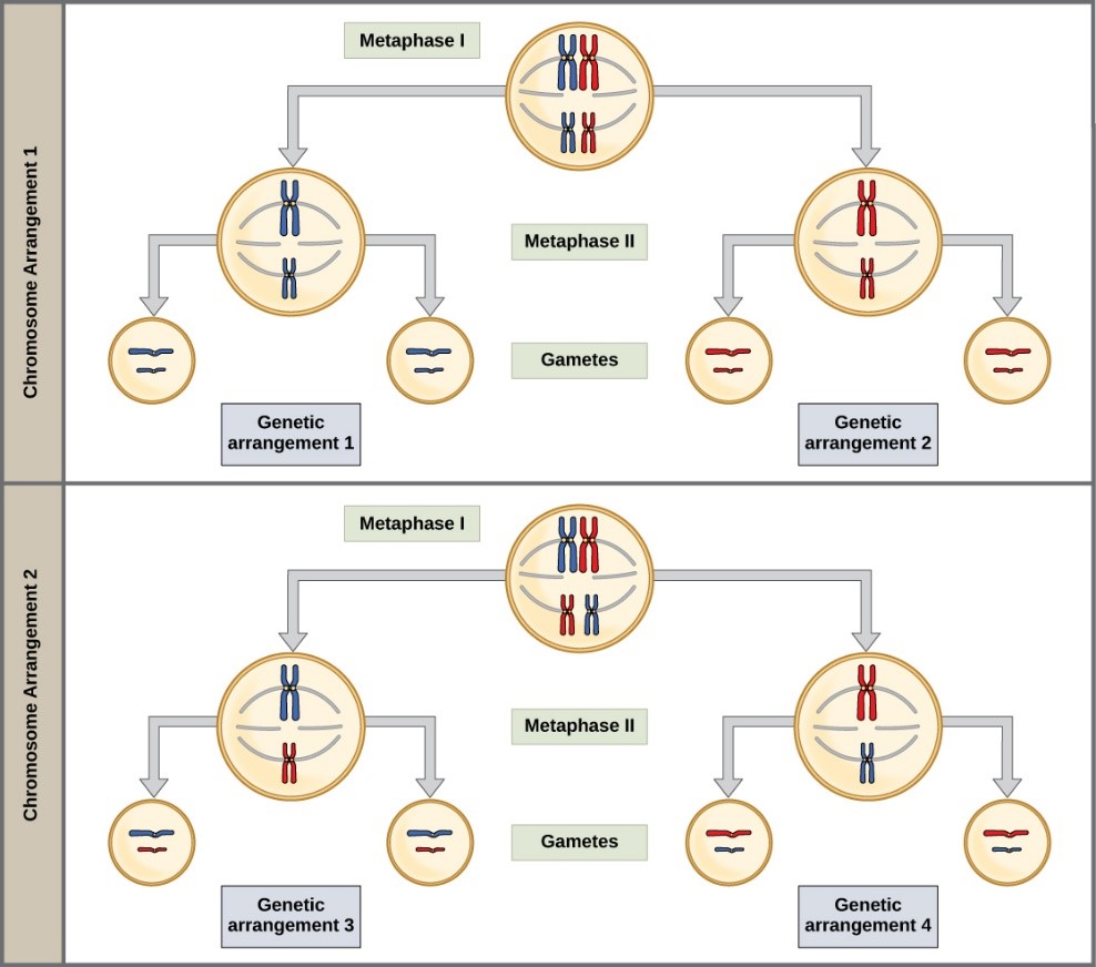
During metaphase I, the homologous chromosomes are arranged in the centre of the cell with the kinetochores facing opposite poles. The orientation of each pair of homologous chromosomes at the centre of the cell is random.
This randomness, called independent assortment, is the physical basis for the generation of the second form of genetic variation in offspring. Consider that the homologous chromosomes of a sexually reproducing organism are originally inherited as two separate sets, one from each parent. Using humans as an example, one set of 23 chromosomes is present in the egg donated by the mother. The father provides the other set of 23 chromosomes in the sperm that fertilises the egg. In metaphase I, these pairs line up at the midway point between the two poles of the cell. Because there is an equal chance that a microtubule fibre will encounter a maternally or paternally inherited chromosome, the arrangement of the tetrads at the metaphase plate is random. Any maternally inherited chromosome may face either pole. Any paternally inherited chromosome may also face either pole. The orientation of each tetrad is independent of the orientation of the other 22 tetrads.
In each cell that undergoes meiosis, the arrangement of the tetrads is different. The number of variations depends on the number of chromosomes making up a set. There are two possibilities for orientation (for each tetrad); thus, the possible number of alignments equals 2n where n is the number of chromosomes per set. Humans have 23 chromosome pairs, which results in over eight million (223) possibilities. This number does not include the variability previously created in the sister chromatids by crossover. Given these two mechanisms, it is highly unlikely that any two haploid cells resulting from meiosis will have the same genetic composition (Figure 4.11).
To summarise the genetic consequences of meiosis I: the maternal and paternal genes are recombined by crossover events occurring on each homologous pair during prophase I; in addition, the random assortment of tetrads at metaphase produces a unique combination of maternal and paternal chromosomes that will make their way into the gametes.
In anaphase I, the spindle fibres pull the linked chromosomes apart. The sister chromatids remain tightly bound together at the centromere. It is the chiasma connections that are broken in anaphase I as the fibres attached to the fused kinetochores pull the homologous chromosomes apart (Figure 4.12).
In telophase I, the separated chromosomes arrive at opposite poles. The remainder of the typical telophase events may or may not occur depending on the species. In some organisms, the chromosomes decondense and nuclear envelopes form around the chromatids in telophase I.
Cytokinesis, the physical separation of the cytoplasmic components into two daughter cells, occurs without reformation of the nuclei in other organisms. In nearly all species, cytokinesis separates the cell contents by either a cleavage furrow (in animals and some fungi), or a cell plate that will ultimately lead to formation of cell walls that separate the two daughter cells (in plants). At each pole, there is just one member of each pair of the homologous chromosomes, so only one full set of the chromosomes is present. This is why the cells are considered haploid—there is only one chromosome set, even though there are duplicate copies of the set because each homolog still consists of two sister chromatids that are still attached to each other. However, although the sister chromatids were once duplicates of the same chromosome, they are no longer identical at this stage because of crossovers.
Meiosis II
In meiosis II, the connected sister chromatids remaining in the haploid cells from meiosis I will be split to form four haploid cells. In some species, cells enter a brief interphase, or interkinesis, that lacks an S phase, before entering meiosis II. Chromosomes are not duplicated during interkinesis. The two cells produced in meiosis I go through the events of meiosis II in synchrony. Overall, meiosis II resembles the mitotic division of a haploid cell.
In prophase II, if the chromosomes decondensed in telophase I, they condense again. If nuclear envelopes were formed, they fragment into vesicles. The centrosomes duplicated during interkinesis move away from each other toward opposite poles, and new spindles are formed. In prometaphase II, the nuclear envelopes are completely broken down, and the spindle is fully formed. Each sister chromatid forms an individual kinetochore that attaches to microtubules from opposite poles. In metaphase II, the sister chromatids are maximally condensed and aligned at the centre of the cell. In anaphase II, the sister chromatids are pulled apart by the spindle fibres and move toward opposite poles.
In telophase II, the chromosomes arrive at opposite poles and begin to decondense. Nuclear envelopes form around the chromosomes. Cytokinesis separates the two cells into four genetically unique haploid cells. At this point, the nuclei in the newly produced cells are both haploid and have only one copy of the single set of chromosomes. The cells produced are genetically unique because of the random assortment of paternal and maternal homologs and because of the recombination of maternal and paternal segments of chromosomes—with their sets of genes—that occurs during crossover.
Also known as programmed cell death, is a normal and controlled cellular process where a cell is instructed to die because it is no longer needed or because it may pose a threat to the organism.
Life cycle of a single cell, from its birth until its division into two new daughter cells.
Part of the cell cycle which includes mitosis and cytokinesis. In this phase DNA replicated in the interphase phase is separated and the cell divides.
Reproductive cells that combine during sexual reproduction to form a new organism.
The process by which haploid cells are formed. Consists of one round of chromosome duplication and two rounds of nuclear division.
Entire life cycle of a cell, excluding mitosis.
Part of the cell cycle which includes mitosis and cytokinesis. In this phase DNA replicated in the interphase phase is separated and the cell divides.
‘Deoxyribonucleic acid’. Contains genetic information for cell function, growth and division.
Made of organised and packaged DNA in the form of genes and are found within the cell nucleus.
Found in eukaryotic cells and contains the DNA genome.
Complex of DNA and proteins found in the nucleus of cells, which helps package DNA into a compact, organised structure.
Region of a chromosome where the spindle fibres attach during cell division, ensuring the proper separation of sister chromatids.
Structure composed of microtubules and proteins that forms during cell division to separate chromosomes into the two daughter cells.
Cylindrical organelles made of microtubules that play a key role in cell division by helping to organise the mitotic spindle and in forming structures like cilia and flagella.
Gel-like substance composed of water and dissolved chemicals.
First stage in mitosis and meiosis. Nuclear envelope begins to break down, spindle fibres start to appear, and chromosome condense and are visible.
Complex nuclear membrane that surrounds the nucleus, consisting of two distinct lipid bilayers contiguous with each other.
Small, membrane-bound sacs within cells that transport, store, and digest substances, playing crucial roles in processes like metabolism, secretion, and cell communication.
Organelles near the nucleus of animal cells that serve as the main microtubule-organising centres, playing a crucial role in cell division by forming the mitotic spindle.
Form part of the cytoskeleton to provide support and are composed of beta and alpha tubulin.
Second phase of mitosis and meiosis. Chromosomes continue to condense, kinetochores appear at centromere and kinetochore microtubules attach.
Third phase of mitosis and meiosis, characterised by the linear alignment of sister chromatids in the centre of the cell.
Also known as equatorial plane. Imaginary plate roughly midway between two poles of the cell that occurs during mitosis and meiosis where sister chromatids line up prior to separation.
Fourth phase of mitosis after metaphase. Sister chromatids are pulled apart and each chromatid is now a chromosome.
Final stage of mitosis and meiosis, preceding cytokinesis, characterised by the formation of two new daughter nuclei.
Following the fusion of two haploid cells, a diploid cell is formed and contains two sets of chromosomes.
Chiasmata (singular: chiasma) are the points at which paired chromosomes (homologous chromosomes) physically cross over each other during the process of meiosis.
