16 Key Cell Types and Tissues in the Human Body
In this section
Content in this section includes:
The most common cell shapes in the human body are squamous (flat), cuboidal (cube-like) and columnar (column-like) and these join together to form tissues that can act as barriers, aid signalling and nutrient and gas transfer and also some can confer protection to underlying tissues or organs. The tissues of multicellular, complex animals can be categorised into four primary types: epithelial, connective, muscle and nervous. Each of these tissues have cells that have a specific role to aid in the functioning of the tissue and in the organs. Each cell has a particular orientation (basal side and apical or free side) and normal cells have a uniform shape, size, function and appearance at a specific location and if changes occur to this appearance and function this can indicate pathology, such as cancer.
Epithelial Tissues
Epithelial tissues cover the outside of organs and structures in the body and line the lumens of organs in a single layer or multiple layers of cells. Epithelial cells fit together tightly to support their protective role in the body. This is accomplished as cells are attached to a basement membrane and each cell is connected to the adjacent cells by special attachments (junctions) which can also aid in cell communication (Figure 5.1).
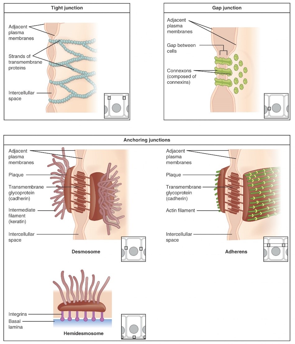
The types of epithelia are classified by the shapes of cells present and the number of layers of cells. Epithelia composed of a single layer of cells attached to a basement membrane is called simple epithelia whereas epithelial tissue composed of multiple layers is called stratified epithelia. Naming of the type of epithelia takes into account the number of cell layers and the cell shape on the free (apical) edge eg a stratified squamous epithelia would have two or more cell layers with squamous cells on the apical (free) surface (Figure 5.2). This architecture also reflects their role/function and location (Table 5.1).
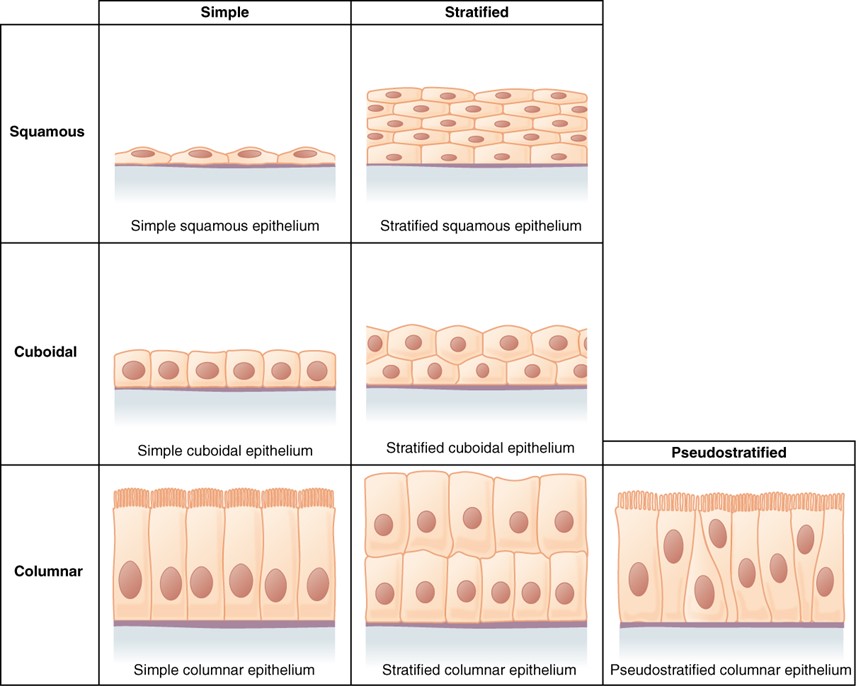
Table 5.1 Types of Epithelial Tissues
| Cell shape | Description | Location | Role/function examples |
|---|---|---|---|
| squamous | flat, irregular, slightly rounded shape | simple: lung alveoli, capillaries; stratified: skin, mouth, vagina | gas exchange in lung alveoli (simple squamous) |
| cuboidal | cube shaped, central nucleus | glands, renal tubules | secretion and absorption in liver and kidneys |
| columnar | tall, narrow, nucleus toward base; tall, narrow, nucleus along cell | simple: digestive tract; pseudostratified: respiratory tract | secretion and absorption in gastrointestinal tract |
| transitional | round, simple but appear stratified | urinary bladder | alows expansion |
Squamous Epithelia
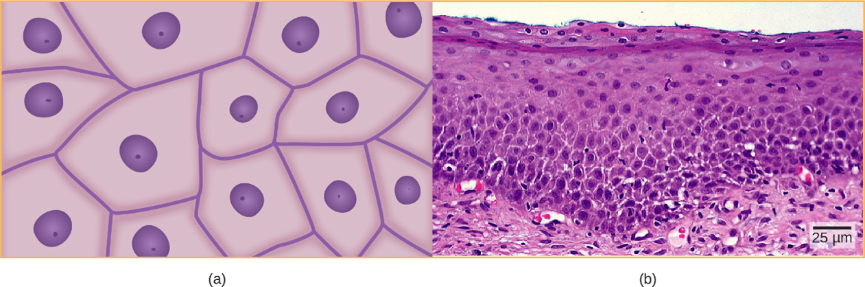
Squamous epithelial cells are generally round, flat and have a small, centrally located nucleus. The cell outline is slightly irregular and cells fit together to form a covering or lining. When the cells are arranged in a single layer on a basement membrane (simple epithelia), they facilitate diffusion in tissues, such as the areas of gas exchange in the lungs and the exchange of nutrients and waste at blood capillaries. When the cells are arranged in multiple layers (stratified epithelia), they form a thicker layer generally used for protection, such as in the skin and in tissues lining the mouth and vagina, where abrasion and damage may occur (i.e. outer cells can be lost but inner cells and membranes are still protected). The human cervix photomicrograph (Figure 5.3b) shows a stratified, squamous epithelium (many layers of cells with squamous cells on the free surface).
Cuboidal Epithelia
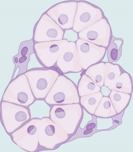
Cuboidal epithelialcells (Figure 5.4), are cube-shaped with a single, central nucleus. They are most commonly found in a single layer (a simple epithelia) and are involved in secretion and absorption depending on the location and need of the cells or tissues. Cuboidal epithelial cells are located in glandular tissues throughout the body, producing and secreting substances such as mucus and digestive juices. These cell types are also found in the walls of tubules and in the ducts of the kidney and liver and play key roles in both secretion and absorption.
Columnar Epithelia
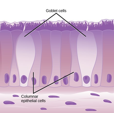
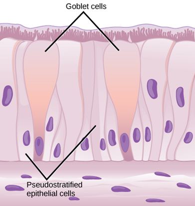
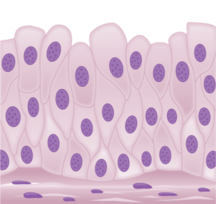
Columnar epithelial cells are taller than they are wide (like a column) and are mostly found as a simple, columnar epithelia with an elongated nucleus located in the basal end of the cell (near the basement membrane; Figure 5.5). This type of epithelia is mostly located in the digestive tract, with the apical surfaces having invaginations (for secretion) and finger-like projections (villi and microvilli) for absorption. Villi and microvilli on the apical surface increase surface area for absorption and therefore increase absorption efficiency of materials from the lumen of the digestive tract then cell prepares it for entry into the body through the circulatory and lymphatic systems. Simple columnar epithelia also line parts of the female reproductive tract and in this location, the apical surface is modified with cilia that beat in unison to aid in the movement of the ovum towards the uterus. Ciliated, columnar epithelia are also found in the respiratory system to help move particulate matter (eg dust particles etc) away from the lungs. Goblet cells (Figure 5.6) are interspersed in some tissues (such as the lining of the trachea). The goblet cells contain mucus that traps irritants, which in the case of the trachea keep these irritants from getting into the lungs.
Transitional Epithelia
Transitional or uroepithelial cells appear only in the urinary system, primarily in the bladder and ureter. These cells are arranged in a stratified layer, but they have the capability of appearing to pile up on top of each other in a relaxed, empty bladder (Figure 5.8). As the urinary bladder fills, the epithelial layer unfolds and expands to hold the volume of urine introduced into it. As the bladder fills, it expands, and the lining becomes thinner – meaning, the tissue transitions from thick to thin.
Connective Tissue
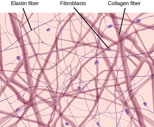
Connective tissues are made up of a matrix consisting of living cells and a nonliving substance, called the ground substance. The ground substance is made of organic substances (usually protein, such as proteoglycan) and an inorganic substance (usually a mineral or water), typically giving a gel-like consistency (with exceptions). The principal cell of connective tissues is the fibroblast. This cell makes the fibres found in nearly all connective tissues. Fibroblasts are motile, able to carry out mitosis and can synthesise whichever connective tissue is needed at that location. Macrophages, lymphocytes and occasionally, leukocytes can be found in some of the tissues. Some tissues have specialised cells that are not found in the others. The matrix in connective tissues gives the tissue its density.
The organic portion or protein fibres found in connective tissues are either collagen, elastic or reticular fibres. Collagen fibres provide strength to the tissue, preventing it from being torn or separated from the surrounding tissues. Elastic fibres are made of the protein elastin; this fibre can stretch to one and one half of its length and return to its original size and shape. Elastic fibres provide flexibility to the tissues. Reticular fibres are the third type of protein fibre found in connective tissues. This fibre consists of thin strands of collagen that form a network of fibres to support the tissue and other organs to which it is connected. There are various types/forms of connective tissue, the types of cells and fibres they are made of, and sample locations of the tissues (Table 5.2).
Table 5.2. Types of connective tissues
| Tissue | Cells | Fibres | Location |
|---|---|---|---|
| loose/areolar | fibroblasts, macrophages, some lymphocytes, some neutrophiles | few: collagen, elastic, reticular | around blood vessels; anchors epithelia |
| dense, fibrous connective tissue | fibroblasts, macrophages | mostly collagen | irregular: skin regular: tendons, ligaments |
| cartilage | chondrocytes, chondroblasts | hyaline: few collagen fibroicartilage: large amount of collagen | shark skeleton, foetal bones, human ears, intervertebral discs |
| bone | osteoblasts, osteocytes, osteoclasts | some: collagen, elastic | vertebrate skeletons |
| adipose | adipocytes | few: collagen, elastic, reticular | adipose (fat) |
| blood | red blood cells, white blood cells | none | blood |
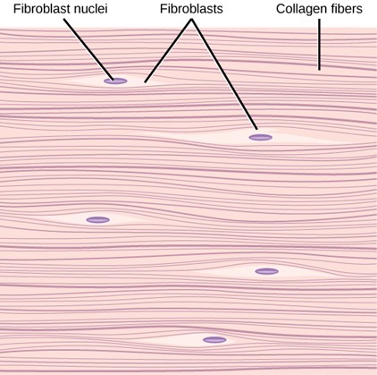
Loose/Areolar Connective Tissue
Loose connective tissue, also called areolar connective tissue, has a sampling of all components of a connective tissue with some fibroblasts together with leukocytes including macrophages, lymphocytes and neutrophils (Figure 5.7). The collagen fibres present are relatively wide and stain a light pink, while elastic fibres are thin and stain dark blue to black. The space between the formed elements of the tissue is filled with the matrix. The material in the connective tissue gives it a loose consistency like a cotton ball that has been pulled apart. Loose connective tissue is found around every blood vessel and helps to keep the vessel in place. The tissue is also found around and between most body organs. In summary, areolar tissue is tough, yet flexible and comprises membranes.
Fibrous Connective Tissue
Fibrous connective tissues contain large amounts of collagen fibres and few cells or matrix material. The fibres can be arranged irregularly or regularly with the strands lined up in parallel. Irregularly arranged fibrous connective tissues are found in areas of the body where stress occurs from all directions, such as the dermis of the skin. Regular fibrous connective tissue (Figure 5.9), is found in tendons (which connect muscles to bones) and ligaments (which connect bones to bones) and due to the dense packing of these fibres in parallel, this gives good resistance to forces along one axis but also allows for a bit of stretch.
Cartilage
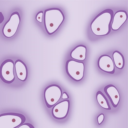
Cartilage is a connective tissue with a large amount of the matrix and variable amounts of fibres. The cells, called chondrocytes, make the matrix and fibres of the tissue. Chondrocytes are found in spaces within the tissue called lacunae.
A cartilage with few collagen and elastic fibres is hyaline cartilage (Figure 5.10). The lacunae are randomly scattered throughout the tissue and the matrix takes on a milky or scrubbed appearance with routine histological stains. Sharks have cartilaginous skeletons, as does nearly the entire human skeleton during a specific pre-birth developmental stage. A remnant of this cartilage persists in the outer portion of the human nose. Hyaline cartilage is also found at the ends of long bones, reducing friction and cushioning the articulations of these bones.
Elastic cartilage has a large number of elastic fibres, giving it tremendous flexibility. The ears of most vertebrate animals contain this cartilage as do portions of the larynx, or voice box. Fibrocartilage contains a large amount of collagen fibres, giving the tissue tremendous strength. Fibrocartilage comprises the intervertebral discs in vertebrate animals. Hyaline cartilage found in movable joints such as the knee and shoulder becomes damaged because of age or trauma. Damaged hyaline cartilage is replaced by fibrocartilage and results in the joints becoming “stiff.”
Bone
Bone, or osseous tissue, is a connective tissue that has a large amount of two distinct types of matrix material. The organic matrix is like the matrix material found in other connective tissues, including some amount of collagen and elastic fibres. This gives strength and flexibility to the tissue. The inorganic matrix consists of mineral salts—mostly calcium salts—that give the tissue hardness. Without adequate organic material in the matrix, the tissue breaks; without adequate inorganic material in the matrix, the tissue bends.
There are three types of cells in bone: osteoblasts, osteocytes, and osteoclasts. Osteoblasts are active in making bone for growth and remodelling. Osteoblasts deposit bone material into the matrix and, after the matrix surrounds them, they continue to live, but in a reduced metabolic state as osteocytes. Osteocytes are found in lacunae of the bone. Osteoclasts are active in breaking down bone for bone remodelling, and they provide access to calcium stored in tissues. Osteoclasts are usually found on the surface of the tissue.
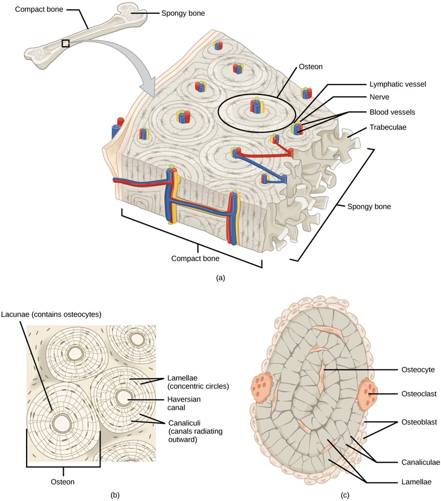
Bone can be divided into two types: compact and spongy. Compact bone is found in the shaft (or diaphysis) of a long bone and the surface of the flat bones, while spongy bone is found in the end (or epiphysis) of a long bone. Compact bone is organised into subunits called osteons (Figure 5.11). A blood vessel and a nerve are found in the centre of the structure within the Haversian canal, with radiating circles of lacunae around it known as lamellae. The wavy lines seen between the lacunae are microchannels called canaliculi; they connect the lacunae to aid diffusion between the cells. Spongy bone is made of tiny plates called trabeculae; these plates serve as struts to give the spongy bone strength. Over time, these plates can break causing the bone to become less resilient. Bone tissue forms the internal skeleton of vertebrate animals, providing structure to the animal and points of attachment for tendons.
Adipose
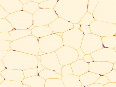
Adipose tissue, or fat tissue, is considered a connective tissue even though it does not have fibroblasts or a real matrix and only has a few fibres. Adipose tissue is made up of cells called adipocytes that collect and store fat in the form of triglycerides, for energy metabolism and also produce a number of growth factors and hormones. Adipose tissues additionally serve as insulation to help maintain body temperatures, allowing animals to be endothermic and they function as cushioning against damage to body organs. Under a microscope, adipose tissue cells appear empty due to the extraction of fat during the processing of the material for viewing (Figure 5.12). The thin lines in the image are the cell membranes and the nuclei are the small, black dots at the edges of the cells.
Blood
Blood is considered a connective tissue because it contains living cells in a matrix (Figure 5.13). The living cell types are erythrocytes (red blood cells (RBC)), and leukocytes (white blood cells (WBC)). The fluid portion of whole blood, its matrix, is commonly called plasma.
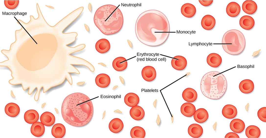
The cell type found in greatest abundance in blood is the erythrocyte. Erythrocytes are consistently the same size in a species but vary in size between species. Mammalian erythrocytes lose their nuclei and mitochondria when they are released from the bone marrow where they are made, whereas fish, amphibian and avian red blood cells maintain their nuclei and mitochondria throughout the cell’s life. The principal job of an erythrocyte is to carry and deliver oxygen to the tissues which is done in all species.
Leukocytes are the white blood cells found in the peripheral blood, with a number of different types with different roles. Lymphocytes function primarily in the immune response to foreign antigens or material. Different types of lymphocytes make antibodies tailored to the foreign antigens and control the production of those antibodies. Neutrophils are phagocytic cells and they participate in one of the early lines of defence against microbial invaders, aiding in the removal of bacteria that has entered the body. Another leukocyte that is found in the peripheral blood is the monocyte. Monocytes give rise to phagocytic macrophages that ‘clean up’ (phagocytose) dead and damaged cells in the body, whether they are foreign or from the host animal. Two additional leukocytes in the blood are eosinophils and basophils — both help to facilitate the inflammatory response.
Platelets or thrombocytes are fragments of a cell made in the bone marrow. Platelets participate in the stages leading up to coagulation of the blood to stop bleeding through damaged blood vessels. Blood has a number of functions, but primarily it transports material through the body to bring nutrients to cells and remove waste material from them while also providing immune functions.
Muscle Tissues
There are three types of muscle in animal bodies: smooth, skeletal and cardiac. They differ by the presence or absence of striations or bands, the number and location of nuclei, whether they are voluntarily or involuntarily controlled and their location within the body (Table 5.3).
Table 5.3. Muscle characteristics
| Type of Muscle | Striations | Nuclei | Control | Location |
|---|---|---|---|---|
| cardiac | yes | single, in centre | involuntary | heart |
| skeletal | yes | many, at periphery | voluntary | skeletal muscles |
| smooth | no | single, in centre | involuntary | visceral organs |
Cardiac Muscle
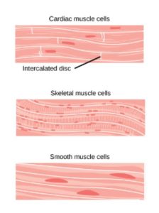
Cardiac muscle (Figure 5.14), is found only in the heart. Like skeletal muscle, it has cross striations in its cells, but cardiac muscle has a single, centrally located nucleus. Cardiac muscle is not under voluntary control but can be influenced by the autonomic nervous system to speed up or slow down. An added feature to cardiac muscle cells is a line than extends along the end of the cell as it abuts the next cardiac cell in the row. This line is called an intercalated disc: it assists in passing electrical impulse efficiently from one cell to the next and maintains the strong connection between neighbouring cardiac cells.
Skeletal Muscle
Skeletal muscle (Figure 5.14), has striations across its cells caused by the arrangement of the contractile proteins, actin and myosin. These muscle cells are relatively long and have multiple nuclei along the edge of the cell. Skeletal muscle is under voluntary, somatic nervous system control and is found in the muscles that move bones.
Smooth Muscle
Smooth muscle (Figure 5.14), does not have striations in its cells. It has a single, centrally located nucleus. Constriction of smooth muscle occurs under involuntary, autonomic nervous control and in response to local conditions in the tissues. Smooth muscle tissue is also called non-striated as it lacks the banded appearance of skeletal and cardiac muscle. The walls of blood vessels, the tubes of the digestive system and the tubes of the reproductive systems are composed of mostly smooth muscle.
Nervous Tissue
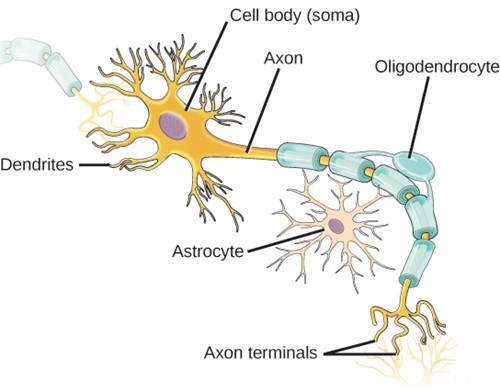
Nervous tissues are made of cells specialised to receive and transmit electrical impulses from specific areas of the body and to send them to specific locations in the body. The main cell of the nervous system is the neuron (Figure 5.15). The large structure with a central nucleus is the cell body of the neuron. Projections from the cell body are either dendrites specialised in receiving input or a single axon specialised in transmitting impulses.
Glial cells (neuroglial cells or glia) are sometimes included in nervous tissue definitions, however they are more of a ‘supportive’ cell type and do not directly engage in electrical signalling but support neurons to carry out this role. There are three distinct types of glial cells: astrocytes (which regulate the chemical environment of the nerve cell); oligodendrocytes (which insulate the axon with myelin sheaths so the electrical nerve impulse is transferred more efficiently) and microglial cells (which are clear cell debris).
Also known as epithelium, it is one of the four main types of tissue in animals. It covers surfaces both internally and externally.
Thin, fibrous, extracellular matrix of tissue that separates the lining of an internal or external body surface (such as epithelial or endothelial tissue) from underlying connective tissue.
Single layer of cells.
Multiple layers of cells staggered on top of one another.
Flat, irregular, slightly rounded shape of epithelial cells
Cube shaped epithelium cells with a central nucleus.
Tall and narrow shaped epithelium cells, with the nucleus located toward base.
Rounded epithelium cells arranged in simple layers, but appear stratified.
One of the four basic types of tissue found in the body. Serves a "connecting" or supportive role, binding, supporting, protecting, and insulating various other tissues and organs in the body.
Nonliving substance made of organic and inorganic substances, typically giving a gel-like consistency.
Main cell of connective tissue, responsible for producing fibres.
Type of white blood cell in the immune system that play several roles in the body's defence against disease. This name reflects their function as phagocytes, cells that can engulf and digest cellular debris, foreign substances, microbes, cancer cells, and anything else that does not have the types of proteins specific to healthy body cells on its surface, in a process called phagocytosis.
Type of white blood cell that are fundamental to the immune system. They are responsible for immune responses that provide long-term protection against pathogens.
Also known as white blood cells, are a key part of the immune system.
Typically refers to the extracellular matrix (ECM), a three-dimensional network of extracellular macromolecules such as collagen, enzymes, and glycoproteins that provide structural and biochemical support to surrounding cells
Main cell of connective tissue, responsible for producing fibres.
Key structural protein that forms the connective tissue throughout our body, from skin to bones, muscles, tendons, and ligaments.
Type of connective tissue composed of the proteins elastin and fibrillin. These fibres provide strength and flexibility to tissues and allow them to return to their original shape after being stretched or contracted.
Type of connective tissue in the body that is composed of parallel bundles of collagen fibers, which give it high tensile strength, i.e., the ability to resist being pulled apart.
Type of firm, flexible connective tissue found in various parts of the body. It is less rigid than bone but more rigid than muscle, and its primary function is to provide support, but unlike bone, cartilage can bend a bit.
Cartilage cells.
Term used in biology to refer to the small spaces or cavities within the hard matrix of bone and cartilage where cells reside.
Type of connective tissue that provides support and flexibility to different parts of the body. It has a glossy, glass-like appearance when cut and viewed under a microscope.
Also known as yellow cartilage, is named "elastic" because of its high content of elastic fibres, which gives it both flexibility and resilience.
Type of cartilage that contains a high proportion of collagen fibres, which gives it significant tensile strength and rigidity. It's less compressible and more dense than other types of cartilage (hyaline and elastic cartilage), which makes it particularly well-suited for areas of the body that require a high degree of support or must withstand heavy pressure.
Refers to anything related to, composed of, or resembling bone.
Cells responsible for the formation of new bone, a process known as ossification. Osteoblasts secrete a matrix of organic compounds, including collagen, which provides a framework for bone growth.
Mature bone cells that originate from osteoblasts.
Large, multinucleated cells that are responsible for the breakdown and resorption of bone tissue, a process known as bone resorption.
Also known as a Haversian system, is the primary structural unit of compact, or cortical, bone.
Singular: canaliculus, are tiny, microscopic channels or passageways in the bone matrix that connect lacunae, the small cavities that house the bone cells (osteocytes), to each other and to the central Haversian canal in an osteon.
Lattice-like network of bony spicules that form the structural units of spongy (cancellous) bone.
Also known as fat tissue, is a specialised type of connective tissue that serves several functions in the body. Its primary role is to store energy in the form of fat, but it also provides insulation and protection for organs.
Lipid storage cells.
Fat molecule; consists of three fatty acids linked to a glycerol molecule.
Also Triacylglycerol.
Endothermic organisms are those that can regulate their body temperature by producing heat metabolically and by insulating the body to reduce heat loss.
Selectively permeable barrier that separates the interior of a cell from its external environment.
Found in eukaryotic cells and contains the DNA genome
Red blood cells (RBCs)
Liquid component of blood in which the blood cells are suspended in. Primarily consists of water, but also contains other substances such as proteins, hormones, nutrients, etc.
Large, complex organelles in which aerobic cellular respiration occurs in eukaryotic cells. Often referred to as the ‘powerhouse’ of the cell.
Type of white blood cell that are fundamental to the immune system. They are responsible for immune responses that provide long-term protection against pathogens.
Type of white blood cell, or leukocyte, that play a crucial role in the body's immune system. They are the largest type of leukocyte and make up about 2-10% of the total leukocyte population in the human body.
Immune cell that surrounds, ingests and destroys foreign material.
Type of white blood cell, or leukocyte, and are part of the body's immune system. They make up about 1-6% of the white blood cells in the bloodstream and are one of the types of cells classified as granulocytes, along with neutrophils and basophils.
Least common type of white blood cell, or leukocyte, making up less than 1% of the total leukocyte population in the blood. Despite their rarity, basophils play important roles in the immune system, particularly in the response to allergies and parasites.
Also known as thrombocytes, are small, colourless cell fragments in our blood that form clots and stop or prevent bleeding.
Pattern of stripes visible under the microscope, primarily in skeletal and cardiac muscle tissues. These striations are caused by the arrangement of contractile proteins, mainly actin and myosin, within the muscle cells.
Membrane-bound organelle found in most eukaryotic cells, often considered the control center of the cell because it houses the cell's genetic material, DNA (deoxyribonucleic acid).
Specialised structures found between the cells (myocytes) of cardiac muscle tissue. They represent complex junctional complexes that are responsible for linking adjacent cardiac muscle cells together, facilitating the coordinated contraction of the heart.
Actin is a family of globular, multi-functional proteins that form microfilaments, one of the three major components of the cytoskeleton in eukaryotic cells. Actin participates in many important cellular processes, including muscle contraction, cell motility, cell division and cytokinesis, vesicle and organelle movement, cell signaling, and the maintenance of cell junctions and cell shape.
Family of motor proteins found in eukaryotic tissues that is best known for its role in muscle contraction. It functions by interacting with another protein called actin, and together, myosin and actin are responsible for producing the force and movement seen in muscle cells.
Also known as a nerve cell, is the fundamental unit of the nervous system, responsible for transmitting information throughout the body. Neurons are electrically excitable cells that function to process and transmit information through electrical and chemical signals.
Glial cells, or simply glia, are non-neuronal cells found in the central nervous system (brain and spinal cord) and the peripheral nervous system. They play several crucial supportive roles in the nervous system, maintaining homeostasis, forming myelin, and providing support and protection for neurons.
Type of glial cell found in the central nervous system, particularly in the brain and spinal cord. They are the most abundant glial cells in the brain and play a vital role in maintaining the health and proper functioning of the nervous system.
Type of glial cell found in the central nervous system (CNS), including the brain and spinal cord. They play a crucial role in supporting and insulating neurons by forming the myelin sheath.
Microglial cells, or simply microglia, are a type of glial cell located throughout the brain and spinal cord. They are the resident macrophages of the central nervous system (CNS) and play a critical role in immune defence, inflammation, and maintenance of homeostasis within the CNS.
