10 Prokaryotic Cells
In this section
Content in this section includes:
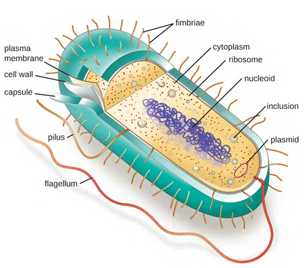
The prokaryotic cell has a relatively simple construction, consisting of a cytoplasm (a gel-like substance composed of water and dissolved chemicals needed for growth), which is contained within a plasma membrane (also called a cell membrane or cytoplasmic membrane); chromosomal DNA, which contains the genetic blueprints of the cell and is concentrated in a nucleoid, ribosomes used to produce proteins and a cell wall. Some prokaryotic cells may also possess flagella, pili, fimbriae and capsules (Figure 3.34). Some other prokaryotic species can produce endospores when conditions are poor for cell growth, and these structures contain all the necessary components to allow the endospore to germinate once it encounters an environment where it can survive and grow. The structures inside a cell are analogous to the organs inside a human body, with unique structures suited to specific functions. A typical prokaryotic cell is illustrated in Figure 3.34.
Common Cell Morphologies and Arrangements
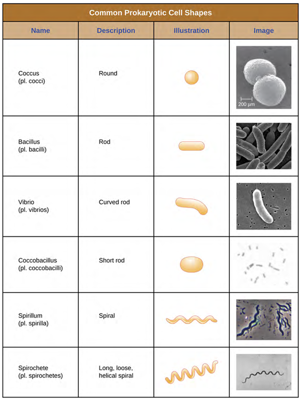
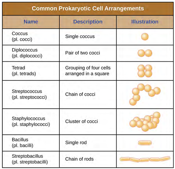
In most prokaryotic cells, morphology is maintained by the cell wall in combination with cytoskeletal elements. Individual cells of a particular prokaryotic organism (species) are typically similar in shape (have a similar cell morphology). Although many thousands of prokaryotic organisms have been identified, only a few different cell morphologies are commonly seen microscopically and include round, rod and spiral shapes (Figure 3.35). In addition to cellular shape, prokaryotic cells of the same species may group together in certain common and distinctive arrangements depending on the plane of cell division (Figure 3.36) and this aids in presumptive identifications.
Nucleoid
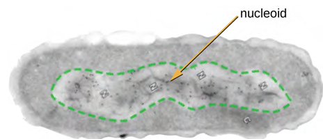
All cellular life has a DNA genome organised into one or more chromosomes. Prokaryotic chromosomes are typically circular, haploid (unpaired) and not bound by a complex nuclear membrane. Prokaryotic DNA and DNA-associated proteins are concentrated within the nucleoid region of the cell (Figure 3.37). In general, prokaryotic DNA interacts with nucleoid-associated proteins (NAPs) that assist in the organisation and packaging of the chromosome. In bacteria, NAPs function like histones, which are the DNA-organising proteins found in eukaryotic cells. In archaea, the nucleoid is organised by either NAPs or histone-like DNA organising proteins.
Photosynthetic Membrane Structures
Some prokaryotic cells, namely cyanobacteria and photosynthetic bacteria, have membrane structures that enable them to perform photosynthesis. These structures consist of an infolding of the plasma membrane that encloses photosynthetic pigments such as green chlorophylls and bacteriochlorophylls. In cyanobacteria, these membrane structures are called thylakoids; in photosynthetic bacteria, they are called chromatophores, lamellae or chlorosomes.
Glycocalyces
Although the majority of prokaryotic cells have cell walls, some may have additional cell envelope structures exterior to the cell wall, such as glycocalyces and S-layers. A glycocalyx is a sugar coat, of which there are two important types: capsules and slime layers.
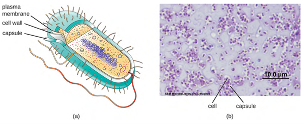
A bacterial capsule is a polysaccharide layer that completely envelopes the cell (Figure 3.38). It is well-organised and tightly packed, which explains its resistance to staining under the microscope. The capsule offers protection from a variety of different threats to the cell, such as desiccation, hydrophobic toxic materials (ie detergents) and bacterial viruses. The capsule can enhance the ability of bacterial pathogens to cause disease and can provide protection from phagocytosis (engulfment by white blood cells known as phagocytes). Lastly, it can help in attachment to surfaces.
The ability to produce a capsule can contribute to a microbe’s pathogenicity (ability to cause disease) because the capsule can make it more difficult for phagocytic cells (such as white blood cells) to engulf and kill the microorganism. Streptococcus pneumoniae, for example, produces a capsule that is well known to aid in this bacterium’s pathogenicity. Capsules are difficult to stain for microscopy and so negative staining techniques are typically used.
A bacterial slime layer is similar to the capsule in that it is typically composed of polysaccharides and it completely surrounds the cell (Figure 3.39). It also offers protection from various threats, such as desiccation and antibiotics. It can also allow for adherence to surfaces. So, how does it differ from the capsule? A slime layer is a loose, unorganised layer that is easily stripped from the cell that made it, as opposed to a capsule which integrates firmly around the bacterial cell wall.

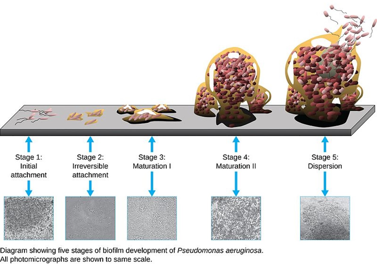
Glycocalyces allow cells to adhere to surfaces, aiding in the formation of biofilms (colonies of microbes that form in layers on surfaces). In nature, most microbes live in mixed communities within biofilms, partly because the biofilm affords them some level of protection. Biofilms generally hold water like a sponge, preventing desiccation. They also protect cells from predation and hinder the action of antibiotics and disinfectants. These properties are advantageous to the microbes living in a biofilm, but they present challenges in a clinical setting, where the goal is often to eliminate microbes. The formation of a biofilm can occur quickly and aids in the colonisation, maturation and then dispersion of certain bacterial populations such as Pseudomonas aeruginosa (Figure 3.40).
S-Layer
Some bacteria have a highly organised layer made of secreted proteins or glycoproteins that self-assemble into a matrix on the outer part of the cell wall. This regularly structured S-layer is anchored into the cell wall, although it is not considered to be officially part of the cell wall in bacteria. S-layers have very important roles for the bacteria that have them, particularly in the areas of growth and survival, and cell integrity.
S layers help maintain overall rigidity of the cell wall and surface layers, as well as cell shape, which are important for reproduction. S layers protect the cell from ion/pH changes, osmotic stress, detrimental enzymes, bacterial viruses, and predator bacteria. They can provide cell adhesion to other cells or surfaces. For pathogenic bacteria they can provide protection from phagocytosis.
Filamentous Appendages
Fimbriae and Pili
Many bacterial cells have protein appendages embedded within their cell envelopes that extend outward, allowing interaction with the environment. These appendages can attach to other surfaces, transfer DNA, or provide movement. Filamentous appendages include fimbriae, pili and flagella (covered earlier).
Fimbriae and pili are structurally similar and because differentiation between the two is problematic, these terms are often used interchangeably. The term fimbriae commonly refers to short bristle-like proteins projecting from the cell surface by the hundreds. Fimbriae enable a cell to attach to surfaces and to other cells. For pathogenic bacteria, adherence to host cells is important for colonisation, infectivity, and virulence. Adherence to surfaces is also important in biofilm formation.
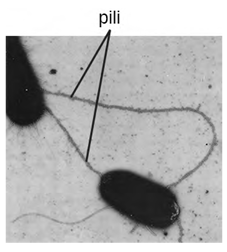
The term pili (singular: pilus) commonly refers to longer, less numerous protein appendages that aid in attachment to surfaces (Figure 3.41). A specific type of pilus, called the F pilus or sex pilus, is important in the transfer of DNA between bacterial cells, which occurs between members of the same generation when two cells physically transfer or exchange parts of their respective genomes, including antimicrobial resistance genes.
Energy Production
The diverse environments and ecosystems on Earth have a wide range of conditions in terms of temperature, available nutrients, acidity, salinity, and energy sources. Prokaryotes are very well equipped to make their living out of a vast array of nutrients and conditions. To live, prokaryotes need a source of energy, a source of carbon, and some additional nutrients.
Cells are essentially a well-organised assemblage of macromolecules and water. Remember, macromolecules are produced by the polymerisation of smaller units called monomers. For cells to build all of the molecules required to sustain life, they need certain substances, collectively called nutrients. When prokaryotes grow in nature, they obtain their nutrients from the environment. Nutrients that are required in large amounts are called macronutrients, whereas those required in smaller, or trace amounts are called micronutrients. Just a handful of elements are considered macronutrients: carbon, hydrogen, oxygen, nitrogen, phosphorus, and sulphur.
Prokaryotes can use different sources of energy to assemble macromolecules from smaller molecules. Phototrophs (or phototrophic organisms) obtain their energy from sunlight. Chemotrophs (or chemosynthetic organisms) obtain their energy from chemical compounds. Chemotrophs that can use organic compounds as energy sources are called chemoorganotrophs. Those that can also use inorganic compounds as energy sources are called chemolithotrophs.
Just as prokaryotes can use different sources of energy, they can also utilise different sources of carbon compounds. The organisms that are able to fix inorganic carbon are called autotrophs and these autotrophic prokaryotes synthesise organic molecules from carbon dioxide. In contrast, heterotrophic prokaryotes obtain carbon from organic compounds. To make the picture more complex, the terms that describe how prokaryotes obtain energy and carbon can be combined. Thus, photoautotrophs use energy from sunlight and carbon from carbon dioxide and water, whereas chemoheterotrophs obtain energy and carbon from an organic chemical source. Chemolithoautotrophs obtain their energy from inorganic compounds, while building their complex molecules from carbon dioxide (Table 3.1).
Table 3.1. Carbon and energy sources in prokaryotes: a summary of the types of energy and carbon sources for different types of prokaryotes to produce energy.
|
Carbon and Energy Sources in Prokaryotes |
||||
|---|---|---|---|---|
|
Energy Sources |
Carbon Sources |
|||
|
Light |
Chemicals |
Carbon dioxide |
Organic compounds |
|
|
Phototrophs |
Chemotrophs |
Autotrophs |
Heterotrophs |
|
|
Organic chemicals |
Inorganic chemicals |
|||
|
Chemo-organotrophs |
Chemolithotrophs |
|||
Source: Table by Boundless, licenced under a CC-BY-SA-4.0 licence.
Archaea and bacteria.
Gel-like substance composed of water and dissolved chemicals.
Selectively permeable barrier that separates the interior of a cell from its external environment.
Structures used by cells to move in aqueous environments – propeller-like.
Gel-like substance composed of water and dissolved chemicals.
Full complement of DNA within a cell organised into smaller, discrete units called genes, arranged on chromosomes and plasmids.
Made of organised and packaged DNA in the form of genes and are found within the cell nucleus.
Archaea and bacteria.
Biological macromolecule comprised of one or more amino acid chains.
Proteins that play a critical role in organising DNA within the nucleus of eukaryotic cells.
Polysaccharide layer that integrates firmly around bacterial cell wall, offers protection to the cell and enhances the cells’ ability to cause disease.
Membranes' major constituent; comprised of two fatty acids and a phosphate-containing group attached to a glycerol backbone.
Immune cell that surrounds, ingests and destroys foreign material.
Ability of an organism, typically a microorganism, to cause disease in another organism.
A colony of bacteria attached to a biotic or abiotic surface enclosed within a matrix of extracellular polymeric substances produced by the colony.
Large, complex molecules that play critical roles in the body, such as assisting in metabolism, transport, stimulation, and cellular replication.
Archaea and bacteria
Large molecule necessary for life that is built from smaller organic molecules
Essential nutrients required for healthy functioning in large quantities.
Essential nutrients required for healthy functioning in small quantities (vitamins and minerals).
Obtain energy from sunlight
Obtain energy from chemical compounds
Chemotrophs that can use organic compounds as energy sources.
Chemotrophs that can use organic and inorganic compounds as energy.
Able to fix inorganic carbon and synthesise organic molecules from carbon dioxide.
Organisms that cannot carbon fix and therefore obtains energy by ingesting other plants or animals.
Generates energy using sunlight and carbon.
Chemotrophs that can use organic and inorganic compounds as energy.
Autotrophs that obtain energy from inorganic compounds.
